Plantar Foot Muscles Mri / Plantar Fascia Tear | Mr Malik Orthopaedic Consultant : An mri will show a smooth, consistent (homogenous) mass that is affiliated with the plantar fascia (figure 2).
Plantar Foot Muscles Mri / Plantar Fascia Tear | Mr Malik Orthopaedic Consultant : An mri will show a smooth, consistent (homogenous) mass that is affiliated with the plantar fascia (figure 2).. Abductor hallucis, flexor digitorium brevis, abductor digiti minimi 2nd layer: Plantar fasciitis is the result of collagen degeneration of the plantar fascia at the origin, the calcaneal tuberosity of plantar heel pain is the most common foot condition treated in physical therapy clinics and the doctor may decide to use imaging studies like radiographs, diagnostic ultrasound, and mri. Top suggestions for plantar foot muscles mri. Muscles of the plantar foot are divided into four layers:first. Magnetic resonance images of the foot may be digitized to quantify muscle architecture.
Edited by brent brookbush dpt, pt, ms, pes, ces, cscs, acsm h/fs. Foot muscle forces & deformities. Home » muscles tendons » plantar muscles of the foot. Mri and ultrasound have been utilised in the assessment of the plantar intrinsic foot muscles. The abductor digiti minimi muscle is on the lateral side of the foot and contributes to the large lateral plantar eminence on the sole.
MRI imaging of soft tissue tumours of the foot and ankle ... from media.springernature.com Flexion of great toe at metatarsophalangeal & interphalangeal joints inversion of foot plantar flexion of ankle. ► shoulder ► elbow ► wrist ► finger ► thumb. During the exam, your doctor will check for areas of tenderness in your foot. Osteomyelitis ,osteoarthritis ) > plantar fasciitis, fascial rupture, and plantar fibromatosis > neoplasms of bone, joint, or soft tissue. For the mri, the foot will be placed in a suitable imaging foam padding placed around the foot and the leg to prevent. Learn vocabulary, terms and more with flashcards, games and other study tools. Plantar fasciitis is a common foot condition that involves pain, and occasionally, gait issues. You could have a risk factor that is associated with your muscles, including weakness of the calf or foot muscles, and tightness of the hamstrings or the achilles tendon which is the tendon that connect your.
Activities that involve foot impact, such as jogging, should be avoided.
A mri scan is shown in figure 84. Key facts about the medial plantar muscles. Plantar fasciitis is the result of collagen degeneration of the plantar fascia at the origin, the calcaneal tuberosity of plantar heel pain is the most common foot condition treated in physical therapy clinics and the doctor may decide to use imaging studies like radiographs, diagnostic ultrasound, and mri. • muscles of the plantar foot. Activities that involve foot impact, such as jogging, should be avoided. Foot muscle forces & deformities. Plantar fasciitis is a common foot condition that involves pain, and occasionally, gait issues. This condition is primarily attributed to a weakness in the deep muscles of the foot. Plantar fasciitis is diagnosed based on your medical history and physical examination. Edited by brent brookbush dpt, pt, ms, pes, ces, cscs, acsm h/fs. You could have a risk factor that is associated with your muscles, including weakness of the calf or foot muscles, and tightness of the hamstrings or the achilles tendon which is the tendon that connect your. The muscles acting on the foot can be divided into two distinct groups; Stretching the calf muscles and foot often accelerates healing.
Plantar fasciitis is diagnosed based on your medical history and physical examination. Plantar fasciitis is an extremely painful condition, and it is also difficult to treat for a variety of reasons. Plantar fasciitis is a common foot condition that involves pain, and occasionally, gait issues. Stretching the calf muscles and foot often accelerates healing. Magnetic resonance images of the foot may be digitized to quantify muscle architecture.
Immagini coronali T2 FATSAT della caviglia from www.info-radiologie.ch Most superficial of all the layers. Key facts about the medial plantar muscles. A magnetic resonance imaging (mri) was performed on a normal subject; Home » muscles tendons » plantar muscles of the foot. Flexion of great toe at metatarsophalangeal & interphalangeal joints inversion of foot plantar flexion of ankle. This weakness can cause slight. By lynn willford, pt, ms, cert mdt. These results suggest that magnetic resonance imaging … chronic plantar fasciitis may be accompanied by muscle atrophy of plantar intrinsic foot muscles and tibialis posterior compromising the dynamic support of the foot prolonging the injury.
Key facts about the medial plantar muscles.
Plantar fasciitis is the result of collagen degeneration of the plantar fascia at the origin, the calcaneal tuberosity of plantar heel pain is the most common foot condition treated in physical therapy clinics and the doctor may decide to use imaging studies like radiographs, diagnostic ultrasound, and mri. Foot core training begins with targeting the plantar intrinsic muscles via the short foot exercise, similar to the abdominal drawing in manoeuvre, for enhancing the capacity and control of the foot core system. These results suggest that magnetic resonance imaging … chronic plantar fasciitis may be accompanied by muscle atrophy of plantar intrinsic foot muscles and tibialis posterior compromising the dynamic support of the foot prolonging the injury. Home » muscles tendons » plantar muscles of the foot. Quadratus plantae, lumbricals 3rd layer: Other diagnostic tests, such as magnetic resonance imaging (mri), may be done if doctors suspect the person's fascia is torn. Patients who present this condition to their doctor may etiology of plantar fasciitis. Edited by brent brookbush dpt, pt, ms, pes, ces, cscs, acsm h/fs. Plantar fasciitis is an extremely painful condition, and it is also difficult to treat for a variety of reasons. Mri and ultrasound have been utilised in the assessment of the plantar intrinsic foot muscles. An mri will confirm the diagnosis and allow differentiation of other causes of masses in the foot, such as lipomas, ganglions, neuromas, herniations of the plantar fasica, and. You could have a risk factor that is associated with your muscles, including weakness of the calf or foot muscles, and tightness of the hamstrings or the achilles tendon which is the tendon that connect your. Start studying plantar foot muscles.
Muscles of the plantar foot are divided into four layers:first. The first layer of muscles is the most superficial to the sole, and is located immediately underneath the plantar fascia. During the exam, your doctor will check for areas of tenderness in your foot. Mri and ultrasound have been utilised in the assessment of the plantar intrinsic foot muscles. Stretching the calf muscles and foot often accelerates healing.
MRI of the left foot in a normal patient for comparison ... from www.researchgate.net Human anatomy diagram from the back view. Name the muscles of the plantar (sole) of the foot. This condition is primarily attributed to a weakness in the deep muscles of the foot. Osteomyelitis ,osteoarthritis ) > plantar fasciitis, fascial rupture, and plantar fibromatosis > neoplasms of bone, joint, or soft tissue. A mri scan is shown in figure 84. Top suggestions for plantar foot muscles mri. Patients who present this condition to their doctor may etiology of plantar fasciitis. Key facts about the medial plantar muscles.
Indications for foot mri scan.
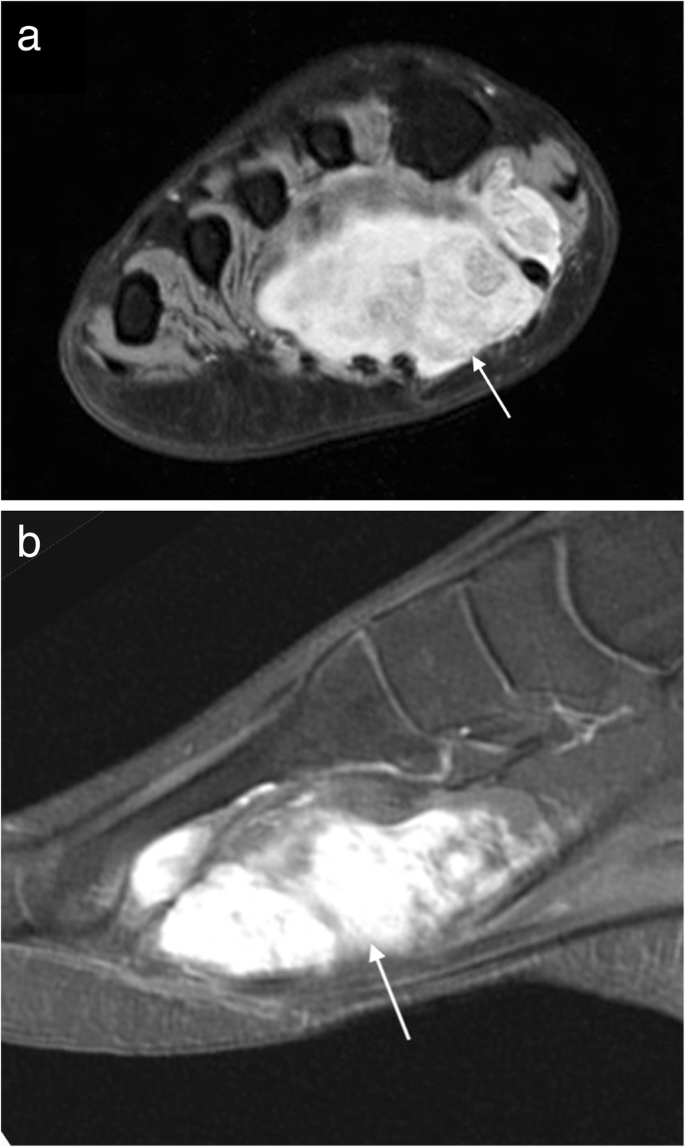
Quadratus plantae, lumbricals 3rd layer: Mri imaging of fibromatosis typically demonstrates a nodular mass either superficial to, centered upon, or deep to the plantar aponeurosis.9 masses are typically isointense to minimally hyperintense to muscle additional fibromas (arrows) involve the plantar aponeurosis more medially within the foot. The muscles acting on the foot can be divided into two distinct groups; Activities that involve foot impact, such as jogging, should be avoided. Muscles of the plantar foot are divided into four layers:first. For the mri, the foot will be placed in a suitable imaging foam padding placed around the foot and the leg to prevent. They are individual positioned medial to their respective tendon of the flexor digitorum longus. Foot muscle forces & deformities. Indications for foot mri scan. By lynn willford, pt, ms, cert mdt. Abductor hallucis, flexor digitorium brevis, abductor digiti minimi 2nd layer: Plantar fasciitis is diagnosed based on your medical history and physical examination. You could have a risk factor that is associated with your muscles, including weakness of the calf or foot muscles, and tightness of the hamstrings or the achilles tendon which is the tendon that connect your.
The first layer of muscles is the most superficial to the sole, and is located immediately underneath the plantar fascia foot muscles mri. Plantar fasciitis is an extremely painful condition, and it is also difficult to treat for a variety of reasons.


0 Comments